External Pacing Technology
External pacing is another term for transcutaneous pacing (TCP), a technology used to treat some forms of arrhythmia. This treatment allows medical personnel to temporarily pace a heart by delivering controlled pulses of electric current.
A healthy adult heartbeat begins with an electrical impulse and while at rest, typically beats between 60 to 100 times per minute. Any variation in this pattern is called an arrhythmia, or an irregular heartbeat, and can indicate a problem with the heart’s electrical system. When an adult heart at rest beats fewer than 60 times a minute, this may indicate a condition called bradycardia, or a slow heart rate. This abnormal heart rhythm is often treated temporarily by transcutaneous, or external, pacing technology.
Transcutaneous pacing should not be confused with defibrillation. Defibrillation is a non-invasive medical technique used to reset the electrical rhythm of the heart during health events such as sudden cardiac arrest or ventricular fibrillation. External pacing is also a non-invasive measure, but instead of delivering a shock, it delivers pulses to keep the heart beating at a steady, regular rhythm until a more permanent form of pacing can be applied. Pacing is also confused with cardioversion, but the two are not the same.
The Difference between Pacing and Cardioversion
Pacing and cardioversion seem similar, as both involve the use of electricity to correct a patient’s heart rate, but they are quite different. Pacing corrects a slow heart rate by delivering controlled pulses to mimic a desired rhythm. Cardioversion is used to restore a fast and unstable heart rate to its normal beating rate through timed shock delivery.
Cardioversion often refers to “synchronized cardioversion,” which corrects a patient’s heart rate by delivering shocks that are timed with particular points on the QRS complex. When a monitor/defibrillator is instructed to begin synchronized cardioversion, it will listen to the patient’s heartbeat and flag R waves while avoiding T waves. When an R wave is identified by the monitor/defibrillator, medical professionals should verify the placement of the R wave, then instruct their manual monitor/defibrillator to deliver energy. When cardioversion is successful, the patient’s heartbeat will begin beating at a normal rhythm.
For an unstable patient with a slow heart rate that doesn’t respond to dopamine or epinephrine, the appropriate treatment is synchronized pacing rather than cardioversion. During the pacing process, the care team uses their monitor/defibrillator to select a healthier heart rate and the level of energy they’d like to deliver along with the shock. This guides the patient’s heart to beat at that particular rate. When successful, pacing will help a patient’s heart resume a healthy rhythm.
Why Pacing Matters
For well-conditioned athletes and some young adults, a slow heart rate is normal. However, for many people, a slow heart rate, or bradycardia, can prevent the heart from pumping enough oxygen-rich blood to vital organs, including the brain. Lack of oxygenated blood can lead to symptoms such as fatigue, dizziness, frequent fainting episodes, shortness of breath, and can even cause more serious conditions such as heart failure, sudden cardiac arrest, or even death.
The History of External Pacing
Dr. Paul Zoll, a founder of ZOLL® Medical Corporation, is credited with modernizing clinical cardiac pacing. He performed the first external human clinical cardiac pacing procedure using the electrodyne PM-65, an early pacemaker of his own design, in 1952.
After the clinical procedure’s initial success, the development of transcutaneous pacing technology continued to progress. In the 1980s, doctors and engineers had a special interest in minimizing patient discomfort, prompting them to develop large adhesive electrodes and ECG filtering. These advancements made transcutaneous pacing the most popular way of treating symptomatic bradycardia and asystole. Today, the company named after Dr. Zoll leads the way in patented external pacing technology.
How ZOLL External Pacing Works
When a patient suffers from bradycardia or another condition for which external pacing is indicated, electrode pads — connected to a monitor/defibrillator — are positioned on the patient’s chest, often directly in front of the heart (anterior), and on the patient’s back, directly behind the heart (posterior). Before starting the pacing process, the care team should use ECG leads and an ECG monitor to confirm electrical capture. OneStep™ Pacing electrodes and OneStep Complete electrodes integrate ECG leads into the anterior electrode, eliminating the need for separate ECG leads and cable.
Once the electrode pads are attached to the patient, the defibrillator is used to deliver strong electrical impulses. This causes the heart to contract in a controlled manner, helping the patient maintain a target heart rate of between 60 and 80 beats per minute until the care team observes an improvement in pulse, blood pressure, skin color, and temperature. This will verify that mechanical capture has occurred successfully.
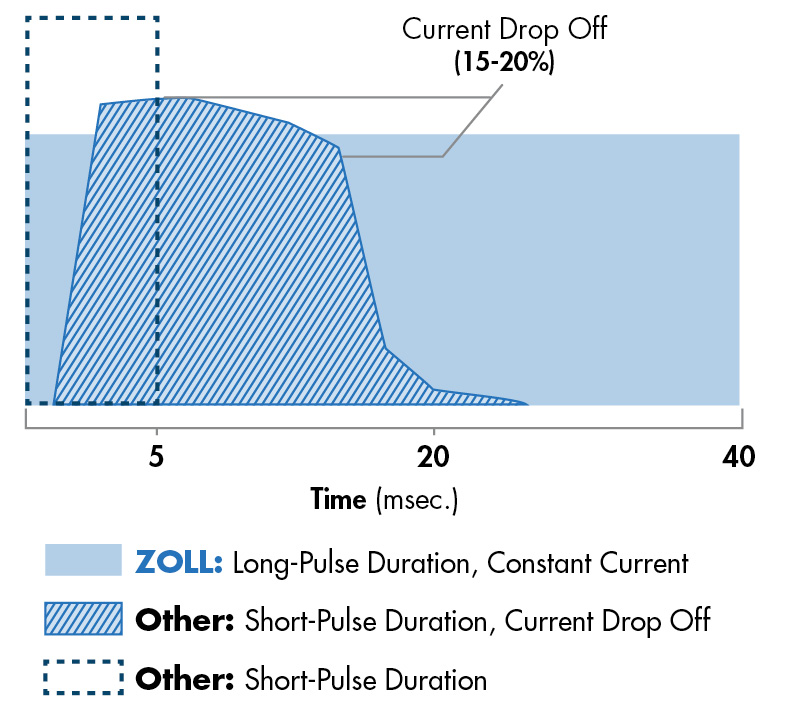
Pacing technology design can greatly impact capture rates and patient survival. The ideal pacer current is the lowest value that maintains capture — usually about 10% above threshold. ZOLL’s patented technology allows ZOLL defibrillators to provide twice the capture of a typical defibrillator at half the current, using a 40-millisecond pulse with constant current. This overcomes disadvantages inherent in other external pacemakers and results in greater patient comfort.
ZOLL also incorporates a unique technology in its R Series® monitor/defibrillator. Pressing and holding the 4:1 button temporarily withholds pacing stimuli, allowing for observation of the patient’s underlying ECG rhythm and morphology without losing capture. When depressed, this button causes pacing stimuli to be delivered at one-fourth of the indicated pulse-per-minute (ppm) setting.

ZOLL’s pacing waveform compared to competitors’
ZOLL helps ensure that a patient’s heart is beating at an acceptable pace with OneStep Pacing electrodes and OneStep Complete electrodes. These electrodes use the ZOLL waveform, which offers the highest rate of capture with the lowest current required.
Both electrode types can be used to help not only with pacing, but also monitoring, defibrillation, and cardioverting. This is accomplished with three different modes: Monitor (color coded with grey), Defibrillation (color coded with red), and Pacing (color coded with green). If you would like to use the electrodes for pacing, change the mode to Pacing and select the proper output and rate for capture.
Applying the electrodes is straightforward, with clear written and illustrative guidance available to the care team. Additionally, the electrodes further simplify the pacing and monitoring process by incorporating ECG electrodes into the anterior electrode, allowing the care team to monitor and pace a patient through a single lead. This inherent feature eliminates the need for a separate ECG cable.
